Microscopy and Image Analysis
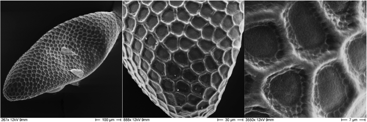
The main focus of the "Microscopic Imaging" department of BNITM is to support projects in molecular parasitology and virology. Another focus is the support of diagnostics in unexplained cases, as well as the detection of alveolar echinococcosis, viral encephalitis and other diseases. For example, electron microscopy was also an important component in the detection of the current Ebola outbreak in March 2014.
The service unit includes the following equipment:
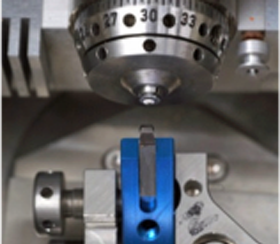
- Transmission electron microscope (120kV Tecnai from FEI) at the LIV
- An ultramicrotome (Leica)
- Sputter coating system

The service unit offers the following techniques:
- Embedding of biological specimen in synthetic resins, plastics
- Preparation of ultrathin sections, also serial sectioning
- 3D reconstruction from serial sections or tomographic datasets
- sputter coating (carbon, glow-discharging)
- Negative contrast imaging of macromolecules, parasites and viruses
- Correlative Light & Electron Microscopy (CLEM)
User
Initially, the employees of the BNITM are considered as users. In individual cases, if the workload of the equipment and staff allows it, employees of other institutions may also use the equipment.
For the planning of the experiments, the users first contact the head of the electron microscopy and arrange an appointment, in which the project is discussed and the experimental procedure is agreed upon.

Contact
- Dr Katharina Scholz-Höhn
- Management Electron microscopy
- phone: +49 40 285380-467
- email: hoehn@bnitm.de


