Research Projects - Parasitology
Sarcocystosis - Outbreak investigations in Malaysia
An outbreak of invasive human sarcocystosis on Tioman Island, Malaysia was epidemiologically investigated, allowing us to describe clinical and immunological characteristics, as well as treatment options. Symptoms, laboratory changes and kinetics of cytokines and chemokines in the patients demonstrated a biphasic course of disease, from an initial vascular to a later muscular stage. Molecular tests for diagnosis of this food-borne parasitosis due to Sarcocystis nesbittii were developed. Local investigations hint at a fecal contamination from large reptiles as source of the outbreak.
Already in the 1990s, sarcocystosis was seen as a possible significant emerging food-borne zoonosis in Malaysia, as an overall seroprevalence of 19.8% was reported among the main racial groups, and 21% of autopsies showed sarcocysts.
In international tourists, the first two waves of the outbreak were associated with travel to Tioman Island mostly during the summer months, while a thrid series of infections was associated with travel in early spring. The course of disease was typically biphasic, with a prodromal stage of one week characterised by fever, myalgia and headache, followed by a two-week asymptomatic period and later by a long-lasting feverish episode with severe myalgia with eosinophilia and CK level elevation. Prolonged and relapsing courses, even over months, have also been documented. Immunologically, a Th2 cytokine polarization and biphasic cytokine changes could be demonstrated.
For a list of the group's publications about sarcocystosis, click here.
Pentastomiasis - Field studies and analysis of local clusters in rural Africa
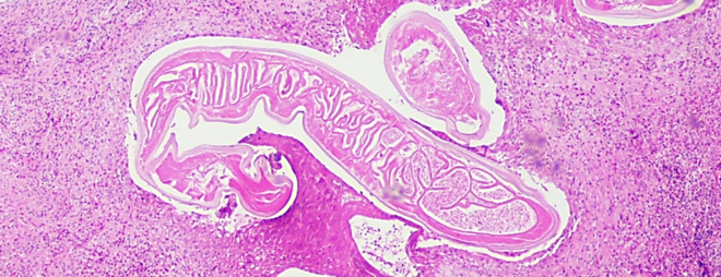
The epidemiology, clinical and histological features of this highly unusual food-borne, tissue-invasive parasitic disease was investigated in two areas of local clustering in rural Africa (The Gambia and the Democratic Republic of The Congo), and Malaysia. Clinically, ocular and visceral forms were detected in two prospective field studies in central Africa. Besides immunohistochemical analyses, specific molecular assays were designed for the detection of parasite larvae even in highly necrotic host tissue. At local markets, Armillifer armillatus and Armillifer grandis pentastomids were discovered in a high percentage of large snakes offered for consumption, emphasizing bushmeat as source of infection with exotic pathogens.
Human infections are caused mainly by larvae of A. armillatus, which is distributed in West and Central Africa. A. grandis, which has drawn recent attention because of heavily symptomatic ocular infections, is prevalent in Central Africa. Two other species, A. moniliformis and A. akgistrodontis, are found in Asia.
Diagnosis relies on radiology and histologic examination. However, determining the pentastomid species, even in nonnecrotic lesions, is nearly impossible and would rely on counting the body annulations in arbitrary tissue section planes. Molecular tools, as shown in our studies, can facilitate diagnosis and are able to determine the causative organism to the species level, also in entirely necrotic and calcified lesions. Even multispecies pentastomid co-infections were found in some patients.
For a list of the group's publications about pentastomiasis, click here.
Echinococcosis & Cysticercosis - Endemic larval tapeworm diseases
Molecular, serological and immunohistochemical assays were engineered for the detection and follow-up analyses of food-borne tissue-invasive larval tapeworm diseases. With a focus on alveolar echinococcosis due to Echinococcus multilocularis, growth characteristics in human tissue were described in a 3D model. In addition, antibody levels against recombinant antigens (especially Em18) were shown to reflect the clinical stage of the disease and to mirror treatment success or relapse, respectively.
Definitive species identification in echinococcosis is of utmost importance, since prognosis and treatment differ fundamentally between the resulting disease forms. Molecular tools, specific immunohistology and serology complement each other to correctly diagnose alveolar or cystic echinococcosis.
For a list of the group's publications about echinococcosis, click here.
Human cysticercosis is commonly caused by the larval stage of the pork tapeworm Taenia solium. Various other zoonotic tapeworm species, which are propagated in different predator-prey cycles, have been described as causes of human cysticercosis, however. Tapeworm larvae have a highly similar morphology, and are histologically often indistinguishable. Molecular methods are able to identify the causative species, and thus the natural reservoir of the respective parasite can be elucidated.
For a list of the group's publications about cysticercosis, click here.
Clinical Parasitology - Nematode Infections
A broad spectrum of various larval nematode infections in humans was clinically, histologically, molecularly and serologically examined. The first German cases of autochthonous Dirofilaria repens infection, Thelazia callipaeda infection, and encephalitic halicephalobiasis were detected and investigated, as well as the first imported infection with Onchocerca lupi into Germany.
Species identification, often only possible with molecular tools, is crucial for prognosis and epidemiological reasons. For a list of the group's publications about nematode infections click here.


